[Product Overview]
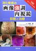
A wealth of case studies are carefully explained using images from endoscopy, CT, MRI, ultrasound, and X-rays.
Includes the anatomy and examination methods of the esophagus, stomach, duodenum, small intestine, and large intestine, with diseases explained on double-page spreads.
This content is useful for everyday practice, so it's recommended for practicing physicians and trainees looking to streamline their knowledge!
[Product Description]
This book focuses on the new era of gastrointestinal imaging diagnosis.
Like other KEY BOOK series books, it covers representative and important diseases and, as a rule, presents them on double-page spreads.
While at first glance it may appear to be a beginner's book, the content is extremely comprehensive.
The book was written by experts in gastrointestinal diagnosis from around the country, primarily from Kumamoto University.
The breadth of cases, the quality of images, and the accuracy of descriptions are unmatched by other books.
One unique feature of this book is that, despite being a book on diagnostic imaging, it actively incorporates endoscopic images, since endoscopic examination is often the first step in treating hollow organs.
It covers the latest imaging methods and important diseases used in gastrointestinal imaging diagnosis, and we believe it is literally the definitive guide to gastrointestinal imaging diagnosis. (Excerpt from the book's preface)
[Author Profile]
Yasuyuki Yamashita
Department of Diagnostic Radiology, Graduate School of Life Sciences, Kumamoto University
<Works>
"KEY BOOK: Imaging Diagnosis of the Liver, Biliary Gland, and Pancreas," "KEY BOOK: Essentials of Urological CT and MRI," "Key Points in Differential Diagnosis of the Liver, Biliary Gland, and Pancreas (Imaging Diagnosis 2016 Special Edition)," and others.
All three are published by Gakken.
[Table of Contents]
1. Esophagus: Normal anatomy and examination of the esophagus, esophageal tumors, esophageal candidiasis and other infections, esophageal achalasia, esophageal hiatal hernia, Plummer-Vinson syndrome, esophageal intramural pseudodiverticulosis
2. Stomach and Duodenum: Normal anatomy and examination of the stomach and duodenum, non-neoplastic gastric lesions, gastric tumors, duodenum
3. Small and Large Intestine: Normal anatomy and examination of the small and large intestine, small intestinal tumors, large intestinal tumors, CT Colonography (CTC), rectum and anus, inflammatory bowel disease (IBD), ischemic enteritis, other enteritis, intestinal diseases, appendix,
vascular lesions, gastrointestinal malignant lymphoma, GIST (gastrointestinal stromal tumor), gastrointestinal neuroendocrine tumors, intestinal endometriosis, gastrointestinal lesions associated with systemic diseases and others,
gastrointestinal varices and ileus, gastrointestinal foreign bodies and complications, volvulus, gastrointestinal perforation, gastrointestinal diverticulum, external hernia, internal hernia, gastrointestinal trauma,
gastrointestinal bleeding, gastrointestinal surgical complications, peritoneum, omentum, abdominal wall, peritoneum, omentum, abdominal wall inflammation, peritoneum, omentum, abdominal wall
4. Pediatrics
Congenital esophageal atresia, tracheoesophageal fistula, hypertrophic pyloric stenosis, duodenal atresia, annular pancreas, intestinal malrotation (mesenteritis commensal) and midgut volvulus,
Hirschsprung Disease, necrotizing enterocolitis, imperforate anus, anorectal malformation
Index